


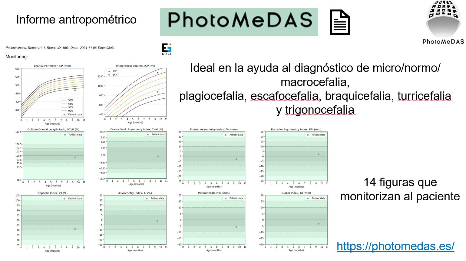
Cranial normality indices in Ugandan infants based on smartphone photogrammetry: key parameters for the study of cranial deformation.
Muñoz-Domingo, M., Romeu-Ferràs, M., Vivas-Consuelo, D., Lerma, J. L., 2026. Head & Face Medicine.
Smartphone 3D scanning technology and 3D semi-synthetic data for processing infant head deformities using artificial intelligence.
Quispe-Enriquez, O. C., Lerma, J. L., 2026. Sensors, 26(5), 1444.
Evaluating the accuracy of smartphone-based photogrammetry and videogrammetry in facial asymmetry measurement.
Teixeira Coelho, L. C., Pinho, M. F. C., Martinez de Carvalho, F., Meneguci Moreira Franco, A. L., Quispe-Enriquez, O. C., Altónaga, F. A., Lerma, J. L., 2025. Symmetry, 17(3), 376.
Craniofacial 3D morphometric analysis with smartphone-based photogrammetry.
Quispe-Enriquez, O. C., Valero-Lanzuela, J. J., Lerma, J. L., 2024. Sensors, 24(1), 230.
Smartphone photogrammetric assessment for head measurements.
Quispe-Enriquez, O. C., Valero-Lanzuela, J. J., Lerma, J. L., 2023. Sensors, 23(21), 9008.
Smartphone-based photogrammetry for craniofacial 3D modelling: A preliminary test.
Quispe-Enriquez, O. C., Valero-Lanzuela, J. J., Baselga, S., Mora-Navarro, G., Lerma, J. L., 2023. Proceedings of 3DBODY.TECH 2023, 14th Int. Conference and Exhibition on 3D Body Scanning and Processing Technologies, 17-18 Oct. 2023, Lugano, Switzerland.
Spherical harmonics to quantify cranial asymmetry in deformational plagiocephaly.
Grieb, J., Barbero-García, I., Lerma, J. L., 2022. Scientific Reports, 12, 167.
Assessment of cranial deformation indices by automatic smartphone-based photogrammetric modelling.
Baselga, S., Mora-Navarro, G., Lerma, J. L., 2022. Applied Sciences, 12(22), 11499.
Combining machine learning and close-range photogrammetry for infant’s head 3D measurement: A smartphone-based solution.
Barbero-García, I., Pierdicca, R., Paolanti, M., Felicetti, A., Lerma, J. L., 2021. Measurement, 182, 109686.
Fully automatic smartphone-based photogrammetric 3D modelling of infant’s heads for cranial deformation analysis.
Barbero-García, I., Lerma, J.L., Mora-Navarro, G., 2020. ISPRS Journal of Photogrammetry and Remote Sensing, 166: 268-277.
Smartphone-based photogrammetric 3D modelling assessment by comparison with radiological medical imaging for cranial deformation analysis.
Barbero-García, I., Lerma, J.L., Miranda, P., Marqués-Mateu, Á., 2019. Measurement, 131: 372-379.
Smartphone-based close-range photogrammetric assessment of spherical objects.
Barbero-García, I., Cabrelles, M., Lerma, J.L., Marqués-Mateu, Á., 2018. The Photogrammetric Record, 33(162): 283-299.
Smartphone-based video for 3D modelling: Application to infant’s cranial deformation analysis.
Lerma, J.L., Barbero-García, I., Marqués-Mateu, Á., Miranda, P., 2018. Measurement, 116: 299-306.
3D photogrammetry in the characterization of deformational plagiocephaly in infants.
Miranda Lloret, P., Garrido García, P., Lerma, J.L., Barbero, I., Plaza Ramirez, E., Alberola, A., 2017. Neurocirugía, 28(Espec Congr): 146.
Syndromic craniosynostosis: photogrammetric evaluation and suturectomy at very early age.
Miranda Lloret, P., Garrido García, P., Lerma, J.L., Barbero, I., Plaza Ramirez, E., Alberola, A., 2017. Neurocirugía, 28(Espec Congr): 394.
Analysis of repeatability on videogrammetry for infants’ cranial deformation.
Barbero-García, I., Lerma, J.L., Marqués-Mateu, Á., Miranda, P., 2017. In: Primer Congreso en Ingeniería Geomática-CIGeo. Martín Furones, Á. (Ed.). Universitat Politècnica de València (pp. 15-19). Julio 5-6, Valencia, Spain.
Low-cost smartphone-based photogrammetry for the analysis of cranial deformation in infants.
Barbero-García, I., Lerma, J.L., Marqués-Mateu, Á., Miranda, P., 2017. World Neurosurgery, 102: 545-554.